Eye Care

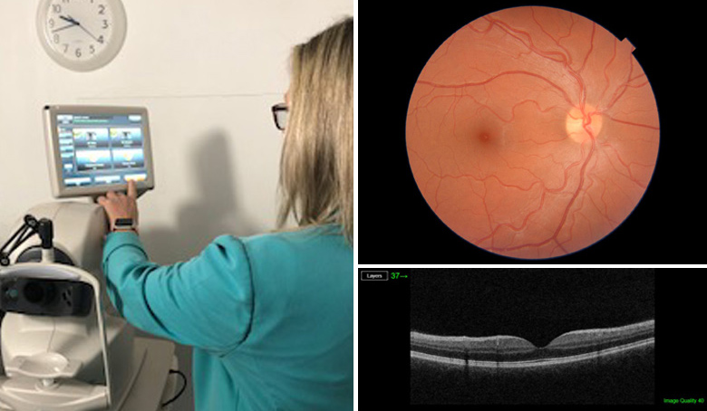
At Scotts Opticians your eye examination will last approximately 45 minutes but sometimes longer. We allow the time to thoroughly examine your eyes and vision using the latest technology such as the OCT scan (featured above). You are encouraged to ask any questions to ensure that you feel at ease. We enjoy testing your eyes and we are very happy to share with you the information we obtain during this process.
It is routine in the practice to screen for all eye conditions including cataracts, macular degeneration, glaucoma and other conditions that may affect the vision. The eye examination at Scotts is personally tailored to you and is based on your needs. You will also be asked to complete a series of Pre-screening tests by our Clinical Assistant and the results are passed onto the Optometrist to aid them in their examination of your eye health and visual status. These preliminary tests include an eye pressure check, field of vision test and a scan of the retina and optic nerve. This is all standard for our private eye examination but there is an additional fee for the scan for the NHS entitled examinations.
The eye examination requires a lot of care and attention. Modern testing equipment helps us to complete the tests accurately, however no equipment on its own is good enough without the skilled knowledge of the Optometrist. We continually strive to have up-to-date knowledge with frequent attendance at continuing education courses and we have good links with the local hospital eye department. We regularly review our testing procedures and make improvements when we can.
Once your examination is complete, the optometrist will explain their findings to you, this includes your eye health and any updates to your prescription that may be required. They will also be happy to answer any questions you have, as well as taking the time to recommend suitable products for your needs and lifestyle and will hand you over to our team of skilled Dispensing Opticians and Dispensing Assistants for frame and lens selection that best suits your needs and style.
Common ocular conditions that can be identified through regular OCT scan screening include:
Age-related macular degeneration (ARMD), diabetes, glaucoma, vitreous detachments and macular holes.
ARMD – is the leading cause of blindness in the UK and can be seen to run in families. It causes gradual deterioration in central vision as the condition affects the macula region of the retina (central portion) which enables detailed vision. There are two types of ARMD – dry and wet.
Dry ARMD is slowly progressive but may not affect the central vision very much in the early stages. Currently there is no treatment for the dry type of ARMD but regular monitoring of the condition is essential as there is a risk of conversion to the wet form.
Wet ARMD causes rapid reduction in central vision and must be treated at hospital very quickly to preserve vision. An injection into the eye may be given by the hospital at regular ongoing intervals to help stabilize the condition.
The OCT scan can help identify the earliest signs of ARMD, to determine if it is the dry or wet form and help monitor its progress over time.
DIABETES – Millions of people around the world have diabetes but there are a significant number of people in the UK with undiagnosed type 2 diabetes (late onset). Diabetic retinopathy is one of the leading causes of blindness in people of working age within the UK.
The OCT scan helps enable early referral and management which can greatly improve the success rate of treatment.
GLAUCOMA – is a condition which causes damage to the optic nerve and causes gradual loss in peripheral vision. The risk of developing glaucoma rises significantly in people over 75 years but it can be seen to run in families and affect them at a younger age. Because the early stages of chronic glaucoma do not cause symptoms, regular eye examinations are essential to diagnose glaucoma at its earliest stage.
The OCT scan can measures features of the optic nerve and look at any changes over time which will help in the early detection of glaucoma.
VITREOUS DETACHMENTS – are usually age-related where the vitreous jelly that takes up the space in the eyeball, decides to separate from the retinal surface. The jelly becomes less firm and can move away from the back of the eye towards the centre. In some cases, parts of the jelly do not detach and cause ‘pulling’ of the retinal surface.
The OCT scan can easily diagnose vitreomacular traction and provide invaluable information about the current relationship between the vitreous and the retinal surface.
MACULAR HOLE – is a small hole in the macula which is at the centre of the retina. A macula hole can affect our ability to read, sew or use a computer. It can form during a complicated vitreous detachment, when the vitreous pulls away from the back of the eye, causing a hole to form. This condition will need to be treat at hospital.
The OCT scan can easily identify if there is a hole in the macula due to the cross sectional view it gives.


